

Image Isn’t Everything…but sometimes it is
When most people think about going to the dentist, they picture cleanings, fillings, maybe whitening. But behind the scenes, there’s a whole world of imaging technology that helps the Chapter One dental team understand what’s really going on with your teeth, gums, and jaw. Think of it as the “backstage pass” to your oral health. And honestly? It’s pretty cool. Let’s break down the main types of imaging you might experience — and why they matter.

What does dental imaging mean for you?
X‑Rays: The Classic (But Way More Advanced Now)
X‑rays are the OG of dental imaging. They’ve been around forever, but today’s digital versions are faster, clearer, and use less radiation. Dental X-rays are recommended for children and adults alike and at Chapter One we are mindful of the extensive guidelines on recommendations surrounding these important diagnostic tools.
What they help us see
Cavities hiding between teeth
Bone levels (important for gum health and implants)
Infections that aren’t visible yet
How teeth are developing in kids and teens
Digital X‑rays pop up on the screen instantly, which means your dentists can walk you through what they’re seeing in real time. It’s a simple, quick way to get a baseline of what’s happening under the surface. With the latest in technology, we can create images that give small detailed insights to localised areas or larger OPGs creating full mouth images. We’ve also fortunately got special cephalometric radiographs that will aid in planning for orthodontics. This isn’t something that is widely available but our specialist orthodontists will love us for - more about that later….
Cone Beam CT (CBCT) Scans: The 3D Upgrade
If X‑rays are the classic movie, CBCT scans are the IMAX version. They create a full 3D image of your teeth, jaw, nerves, and bone structure.
Why CBCT is a game‑changer
Implant planning becomes incredibly precise
Nerve locations are mapped out clearly
Sinus anatomy is visible for more complex treatments
Root canals can be planned with more accuracy
This is the kind of imaging that helps dentists plan treatments down to the millimetre. It’s especially helpful if you’re getting implants or any advanced restorative work. We can use the CBCT image - a 3d insight into your anatomy to plan aesthetic treatments from the outside in. Making procedures more predictable and accurate. Dr Chris has gone through special training to be able to assess and translate these images all for your benefit.
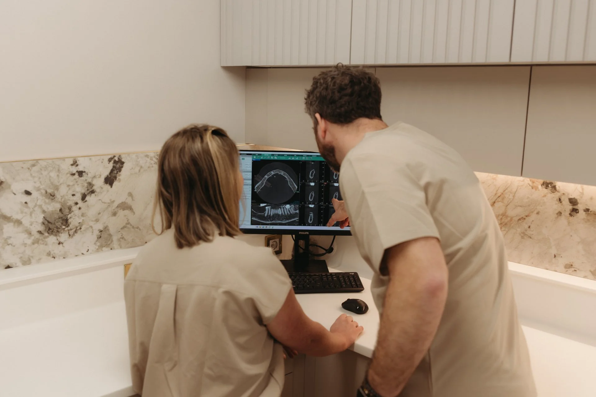
Dr Ashley and Dr Chris taking the time to review slices through a 3D CBCT scan
Intra‑Oral Scanners: Goodbye Goopy Impressions
If you’ve ever had traditional dental impressions, you probably remember the trays, the putty, and the urge to gag. Intra‑oral scanners have basically made all of that unnecessary. We can use the latest in technology to create a 3d model of your mouth without messy impressions that can often create inaccuracies.
Why patients love digital scanning
No mess, no discomfort
Super accurate 3D models
Instant visuals you can see on the screen
Faster turnaround for crowns, aligners, and other treatments
Your dentist simply moves a small wand around your mouth, and a digital model appears like magic. It’s one of the easiest, most patient‑friendly pieces of tech in dentistry today.
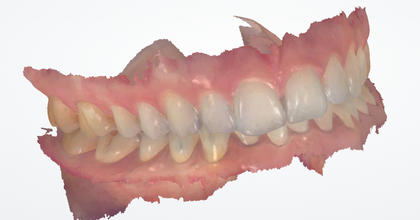
Your teeth in a 3d digital model
Intra‑Oral & Extra‑Oral Photos: The Storytelling Tools
Photos might seem simple, but they’re incredibly powerful. They help document your smile, track changes, and plan cosmetic or restorative treatments. Sometimes its a patient’s first insight into their own mouths. We love digital photography and as part of the Chapter One family, you will too!
Intra‑oral photos
These are the close‑ups taken inside your mouth. They show:
Tooth colour and texture
Gum health
Wear and tear
Existing restorations
Recessions and cracks
Extra‑oral photos
These are taken from the outside — your smile, your profile, your facial symmetry. They’re especially helpful for:
Smile design
Cosmetic planning
Before‑and‑after comparisons
Together, these photos help your dentist see the full picture — not just what’s happening tooth‑by‑tooth, but how everything fits your face and personality.
How It All Comes Together
Each type of imaging gives a different piece of the puzzle. When you combine them, you get a complete, detailed understanding of your oral health.
This helps your dental team:
Explain things clearly
Catch issues early
Plan treatments more precisely
Create results that look natural and last longer
We love the transparency of letting you be fully engaged in your own health
It also helps you feel more informed and confident about your care. We love creating images to engage you with your own oral health and we won’t stop keeping up with the latest in dental imaging technology so you don’t have to.
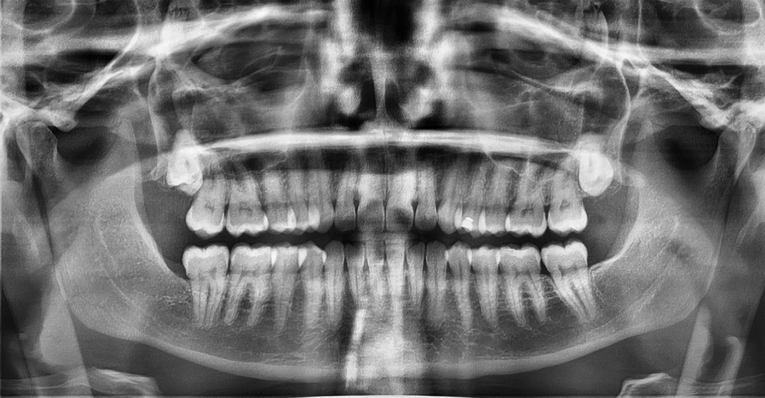
A full OPG radiograph of your mouth
What a Typical Visit Might Include
Depending on your needs, you might experience a mix of:
Digital X‑rays for a quick health check
CBCT scan if you’re planning implants or complex work
Intra‑oral scanning for digital models
Photos to document and design your smile
Everything is shown to you on screen so you can actually see what’s going on — no guessing, no confusion.
The Future of Dental Imaging
Technology keeps evolving, and dentistry is evolving with it. We’re seeing:
Even lower‑dose imaging
AI‑assisted diagnostics
More advanced 3D modelling
Better integration between scanners, labs, and treatment planning
But at the end of the day, the goal stays the same: to understand your smile better and give you care that feels personal, precise, and genuinely thoughtful. We are so excited to be one of a handful of dental practices in Edinburgh offering Pearl dental AI assisted diagnostics on our radiographs allowing our clinicians not only to read radiographs but to have assistance in communication and diagnosis of findings from the latest in dental technology.
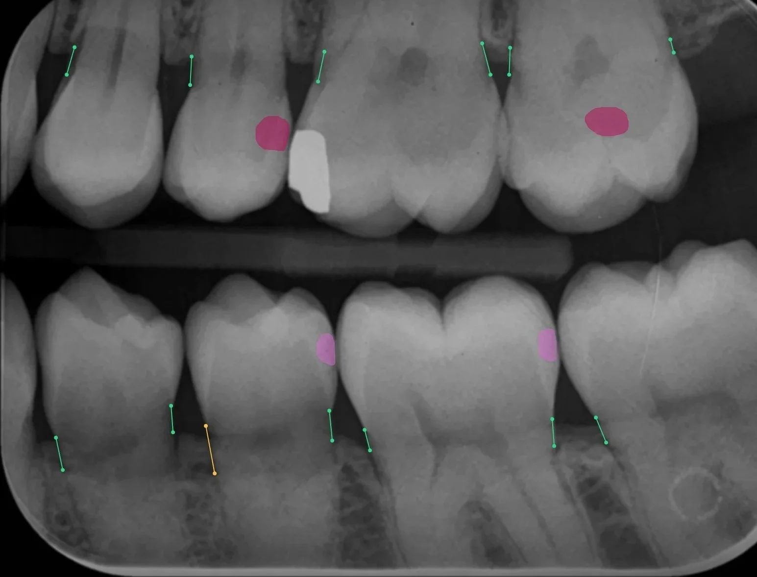
How Pearl Dental AI can assist in diagnostic imaging
A Quick Shoutout: Our Nurses Are Levelling Up
Here’s something we’re genuinely excited about: Our Chapter One nurses are currently training to become radiography‑qualified. We’re delighted to be levelling up as all of our staff get on board this exciting field of dentistry.
That means even more of our team will be able to take X‑rays and support advanced imaging — safely, confidently, and with the same thoughtful approach we bring to everything else.
It’s a big step, and we’re incredibly proud of them. They’re already brilliant, and this just adds another superpower to their toolkit.
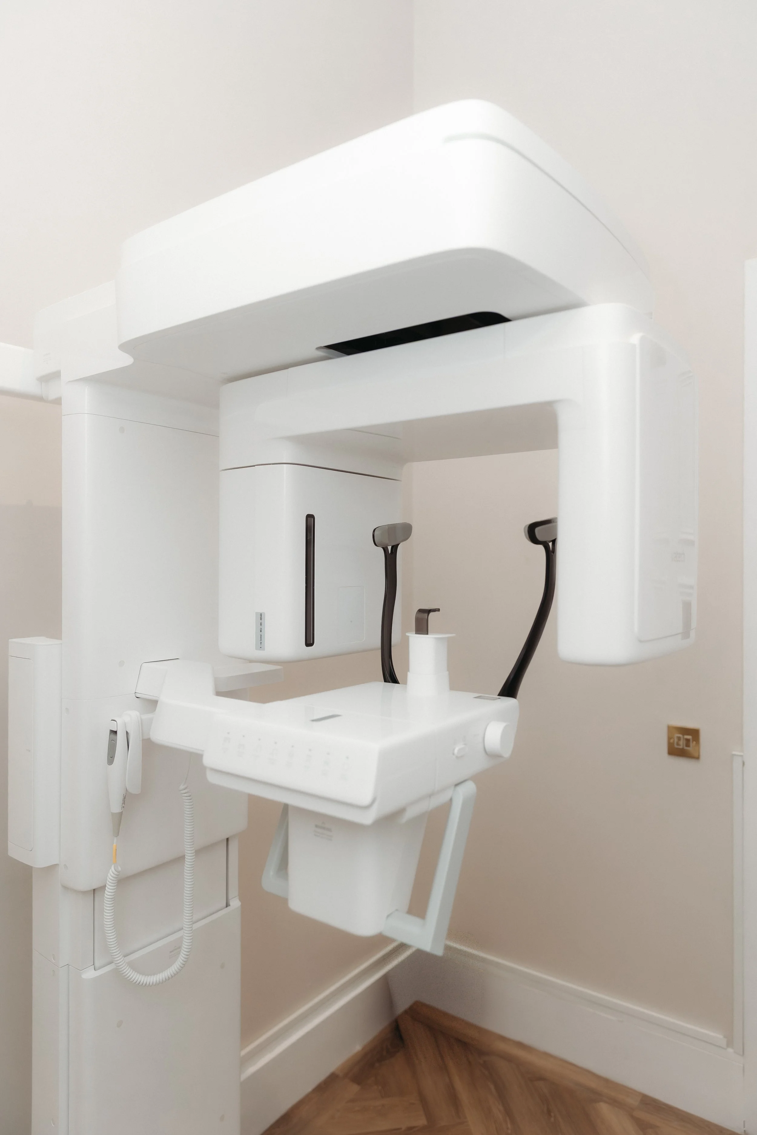
Vatech CBCT and cephalometric scanner
Why Chapter One Dental?
Our clinic brings every corner of modern dental imaging under one roof, making your visit smoother, smarter, and surprisingly fun. With crystal‑clear clinical photography, precision radiography, detailed 3D scans, advanced CBCT imaging, and the sharp eye of Pearl AI, we turn complex technology into simple confidence.
No more bouncing between providers or guessing what comes next — everything you need is right here, working together seamlessly. When your smile deserves the best tools in the business, why settle for anything less than a one‑stop shop that has them all? Because at Chapter One Dental, our story starts with your smile. 🦷✨